BACKGROUND – The routine acquisition of combined prone and supine SPECT myocardial perfusion images remains limited despite the associated improvement in diagnostic and prognostic accuracies. Our aim was to determine whether the assessment of regional wall thickening (WT) in addition to myocardial perfusion from stress supine acquisitions could overcome the lack of prone acquisition and the corresponding decrease in the diagnostic performances of SPECT myocardial perfusion imaging (MPI) in patients with known or suspected coronary artery disease (CAD).
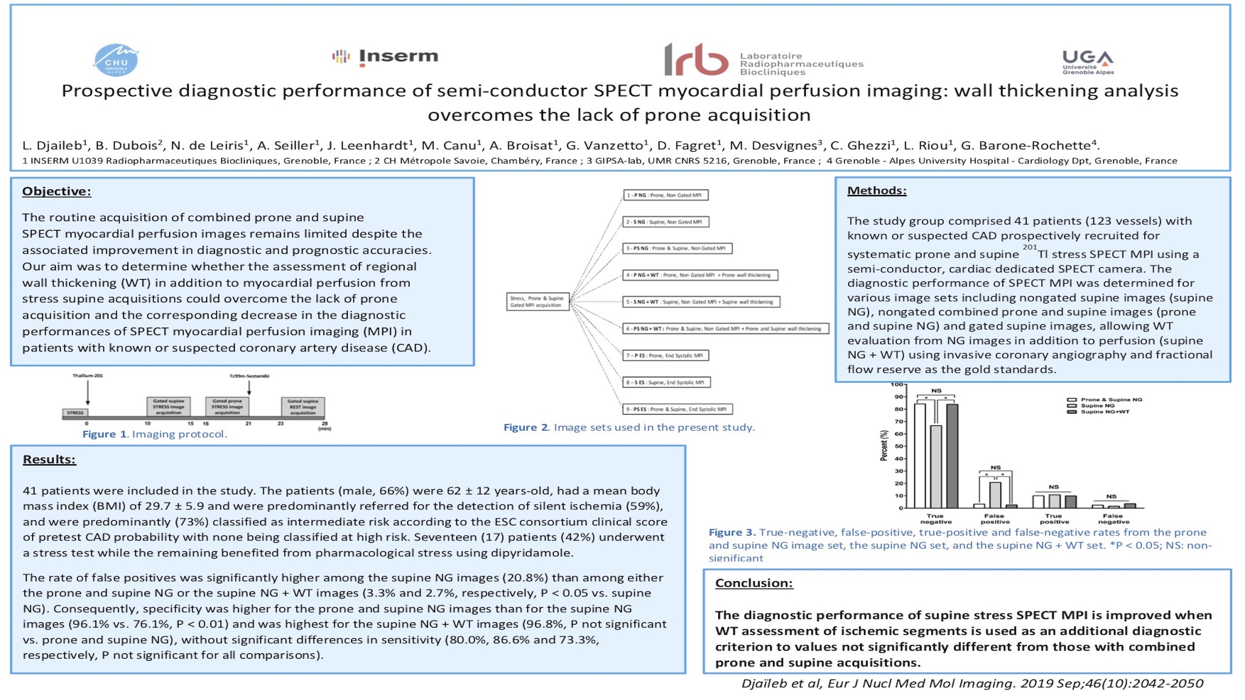
METHODS – Forty-one (41) patients (123 vessels) with known or suspected CAD were prospectively recruited for systematic prone and supine thallium-201 stress SPECT myocardial perfusion imaging using a semi-conductor, cardiac dedicated SPECT camera. The diagnostic performances of SPECT MPI were determined for various image sets including non-gated, supine images (supine NG), non-gated, combined prone and supine acquisitions (prone & supine NG), and gated supine acquisition allowing the evaluation of WT in addition to that of perfusion from NG images (supine NG+WT) using invasive coronary angiography (ICA) and fractional flow reserve (FFR) as the gold standard. Myocardial perfusion was scored using a 5-point scale (0 = normal, 1 = equivocal, 2 = moderate, 3 = severely reduced radiotracer uptake, 4 = no detectable uptake) and a 17-segment model of the left ventricle leading to summed stress score values (SSS), with patients being classified as positive for the presence of ischemia if SSS>=2. Additional WT analysis was used to classify myocardial perfusion as pathological when a myocardial segment with a SSS>=2 also presented with a WT abnormality. Significant CAD was defined by the presence of >=90% stenosis/occlusion or fractional flow reserve <= 0.80 in the presence of a >=50% coronary stenosis.
RESULTS – Upon per patient analysis, the occurrence of false positives was significantly higher in supine NG images (36.6%) than in both prone & supine NG and supine NG+WT image sets (9.8% and 7.8%, respectively, p<0.05 vs supine NG) (see Figure). Consequently, specificity decreased from prone & supine NG to supine NG images (86.2% vs. 48.3%, p=0.003) and was restored when using supine NG+WT images (88.5%, p=NS vs prone & supine NG), with no compromise in sensitivity (91.7%, 91.7%, 83.3%, respectively, p=NS for all comparisons). Similar results were observed upon per vessel analysis.
CONCLUSIONS - The diagnostic performances of supine stress SPECT MPI are restored compared with combined prone & supine acquisitions when WT assessment in the ischemic segments is used as an additional diagnostic criterion.